Achilles tendon
| Achilles tendon | |
|---|---|
 |
|
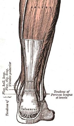
| Posterior view of the foot and leg, showing the Achilles tendon (tendo calcaneus). The gastrocnemius muscle is cut to expose the soleus. | |
| Lateral view of the human ankle, including the Achilles tendon | |
| Latin | tendo calcaneus, tendo Achillis |
| Gray's | subject #129 483 |
| MeSH | Achilles+tendon |
The Achilles tendon (or occasionally Achilles’ tendon), also known as the calcaneal tendon or the tendo calcaneus, is a tendon of the posterior leg. It serves to attach the plantaris, gastrocnemius (calf) and soleus muscles to the calcaneus (heel) bone.
Contents |
Anatomy
The Achilles is the tendonous extension of two muscles in the lower leg: gastrocnemius and soleus . In humans, the tendon passes behind the ankle. It is the thickest and strongest tendon in the body. It is about 15 centimetres (5.9 in) long, and begins near the middle of the calf, but receives fleshy fibers on its anterior surface, almost to its lower end. Gradually becoming contracted below, it is inserted into the middle part of the posterior surface of the calcaneus, a bursa being interposed between the tendon and the upper part of this surface. The tendon spreads out somewhat at its lower end, so that its narrowest part is about 4 centimetres (1.6 in) above its insertion. It is covered by the fascia and the integument, and stands out prominently behind the bone; the gap is filled up with areolar and adipose tissue. Along its lateral side, but superficial to it, is the small saphenous vein. The Achilles' muscle reflex tests the integrity of the S1 spinal root. The tendon can receive a load stress 3.9 times body weight during walking and 7.7 times body weight when running.[1]
Nomenclature
The oldest-known written record of the tendon being named for Achilles is in 1693 by the Flemish/Dutch anatomist Philip Verheyen. In his widely used text Corporis Humani Anatomia, Chapter XV, page 328, he described the tendon's location and said that it was commonly called "the cord of Achilles" ("quae vulgo dicitur chorda Achillis").
The name Achilles' heel comes from Greek mythology. Achilles' mother, the goddess Thetis, received a prophecy of her son's death. Hearing this, she dipped him into the River Styx to protect his body from harm. However, she kept hold of his heel, meaning that the water did not touch this part of his body and it was therefore vulnerable. During the Trojan War, Achilles was struck on his unprotected heel by a poisoned arrow, which killed him. In the same war, Achilles is also said to have cut behind Hector's Achilles tendons, having killed him, and threaded leather thongs through the incisions in order to drag him behind a chariot.
Because eponyms have no relationship to the subject matter, anatomical eponyms are being replaced by descriptive terms. The current terminology for Achilles tendon is calcaneal tendon. However, recently the medical community has decided to revert to the old eponyms; and this tendon is, once again, known as the Achilles.
Role in disease
The most common Achilles tendon injuries are Achilles tendinosis and Achilles tendon rupture. Achilles tendinosis is the soreness or stiffness of the tendon, generally due to overuse. Achilles tendinitis (inflammation of the tendon) was thought to be the cause of most tendon pain, until the late 90s when scientists discovered no evidence of inflammation. Partial and full Achilles tendon ruptures are most likely to occur in sports requiring sudden eccentric stretching, such as sprinting. Maffulli et al. suggested that the clinical label of tendinopathy should be given to the combination of tendon pain, swelling and impaired performance. Achilles tendon rupture is a partial or complete break in the tendon; it requires immobilization or surgery. Xanthoma can develop in the Achilles tendon in patients with familial hypercholesterolemia.
Treatment of damage
Initial treatment of damage to the tendon is generally nonoperative. Orthotics can produce early relief to the tendon by the correction of malalignments, non-steroidal anti-inflammatory drugs (NSAIDs) are generally to be avoided as they make the more-common tendinopathy (degenerative) injuries worse; though they may very occasionally be indicated for the rarer tendinitis (inflammatory) injuries. Physiotherapy by eccentric calf stretching under resistance is commonly recommended, usually in conjunction with podiatric insoles or heel cushioning. According to reports by Hakan Alfredson, M.D., and associates of clinical trials in Sweden, the pain in Achilles tendinopathy arises from the nerves associated with neovascularization and can be effectively treated with 1–4 small injections of a sclerosant. In a cross-over trial, 19 of 20 of his patients were successfully treated with this sclerotherapy.
In a case where Achilles tendon rupture is concerned, there are three main types of treatment: the open and the percutaneous operative methods, and nonoperative approaches.
Depending on the severity of the injury, recovery from an Achilles injury can take up to 12–16 months.
Evolution and function
The Achilles tendon is short or absent in great apes, but long not only arboreal gibbons and humans.[2] It provides elastic energy storage in hopping,[3] walking, and running.[2] Computer models suggest this energy storage Achilles tendon increases top running speed by >80% reduces running costs by more than three-quarters.[2] It has been suggested that the "absence of a well-developed Achilles tendon in the nonhuman African apes would preclude them from effective running, both at high speeds and over extended distances."[2]
Role in postural orientation
Bilateral Achilles tendon vibration in the absence of vision has a major impact on postural orientation. [1] Vibration applied to the Achilles tendon is well known to induce in freely standing subjects a backward body displacement and in restrained subjects an illusory forward body tilt. [2] The vibrations stimulate muscle spindles in the calf muscles. The muscle spindles alert the brain that the body is moving forward, so the central nervous system compensates by moving the body backwards.
See also
- Heel lifts
References
- ↑ Giddings VL, Beaupré GS, Whalen RT, Carter DR. (2000). Calcaneal loading during walking and running. Med Sci Sports Exerc. 32(3):627-34. PMID 10731005
- ↑ 2.0 2.1 2.2 2.3 Sellers WI, Pataky TC, Caravaggi P, Crompton RH. (2010). Evolutionary Robotic Approaches in Primate Gait Analysis. Int J Primatol 31:321–338 doi:10.1007/s10764-010-9396-4
- ↑ Lichtwark GA, Wilson AM. (2005). In vivo mechanical properties of the human Achilles tendon during one-legged hopping. J Exp Biol. 208(Pt 24):4715-25. PMID 16326953
Additional images
|
The popliteal, posterior tibial, and peroneal arteries. |
Back of left lower extremity. |
 The mucous sheaths of the tendons around the ankle. Lateral aspect. |
 The mucous sheaths of the tendons around the ankle. Medial aspect. |
|
||||||||||||||||||||||||||||||||||||||||||||||||||||||||